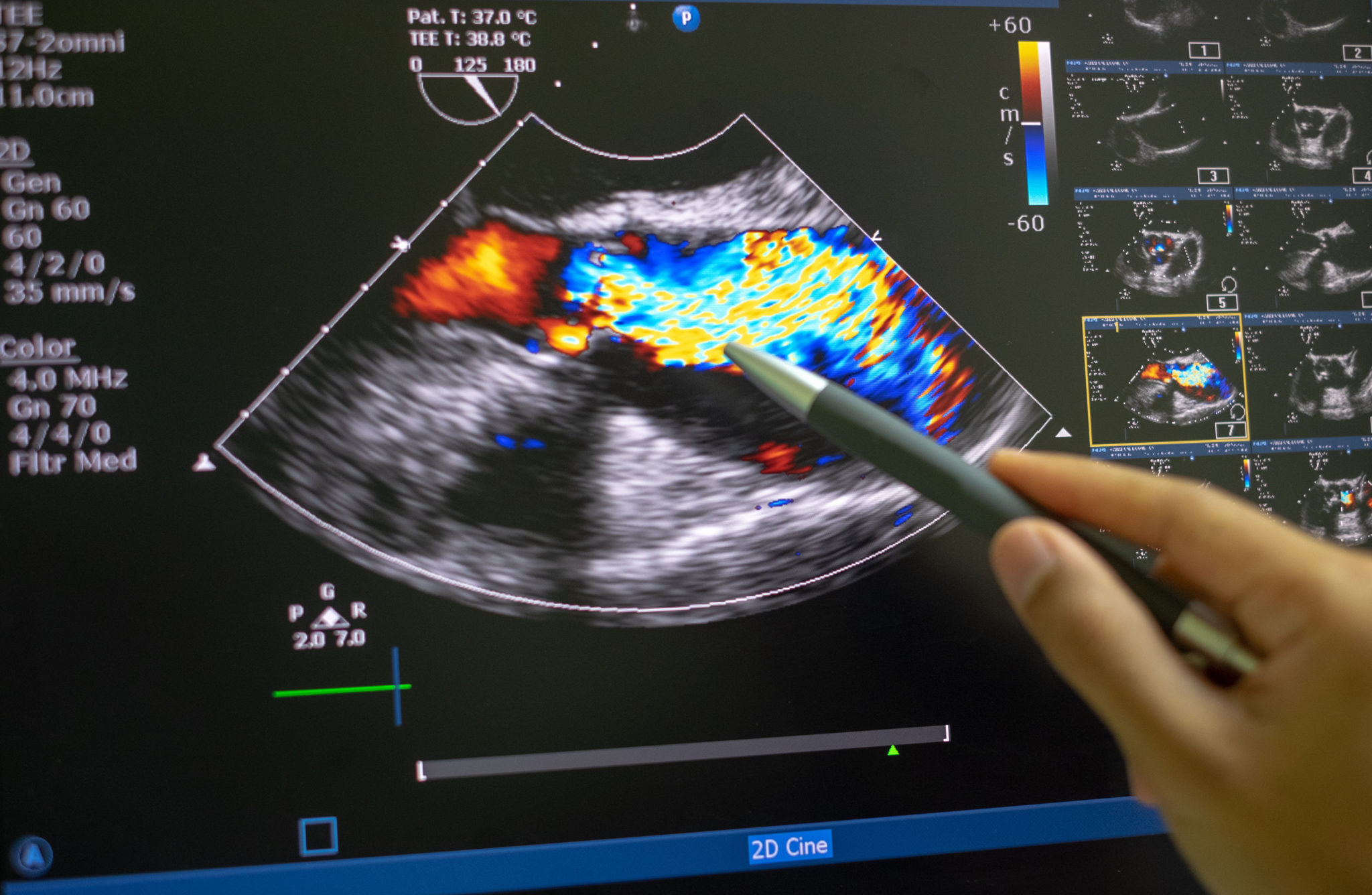
Transesophageal Echocardiography Training for Critical Care Fellows Pulmonology Advisor
Takeaway. Echocardiography is a test using sound waves to produce live images of your heart. The image is called an echocardiogram. It allows your doctor to monitor how your heart and its valves.
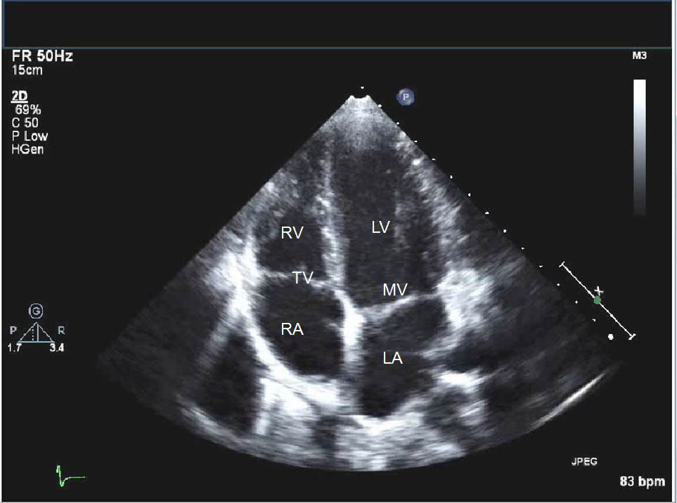
Transthoracic Echocardiogram One Heart Cardiology
Echocardiography, also known as cardiac ultrasound, is the use of ultrasound to examine the heart.It is a type of medical imaging, using standard ultrasound or Doppler ultrasound. The visual image formed using this technique is called an echocardiogram, a cardiac echo, or simply an echo.. Echocardiography is routinely used in the diagnosis, management, and follow-up of patients with any.
What To Expect From An Echocardiogram hubpages
Overview. An echocardiogram is a common test that uses high-frequency ultrasound waves to create a moving picture of the heart while it is beating. It shows the size and shape of the heart, and provides images of the chambers, walls, valves, and blood vessels, tipping off your doctor if there are any problems. Echocardiograms are used to assess.
Echocardiogram Heart Clinic
Echocardiography is the official publication of the International Society of Cardiovascular Ultrasound. As a respected cardiac and cardiovascular imaging journal, Echocardiography is recognized for its comprehensive peer-reviewed articles, case studies, original research, and reviews by international authors.The journal keeps its readership of echocardiographers, ultrasound specialists, and.

Transthoracic Echocardiography in Australia Dr Arthur Nasis
A 2D echo is the standard test, which shows your doctor images of your heart's walls, valves, and some vessels. If they need more detailed pictures, especially of your heart's lower left chamber.
Echocardiogram
This type of echocardiogram is done during pregnancy to check the baby's heart. It's a noninvasive test that involves moving an ultrasound wand over the pregnant person's belly. It lets a health care provider see the unborn baby's heart without using surgery or X-rays. Stress echocardiogram.
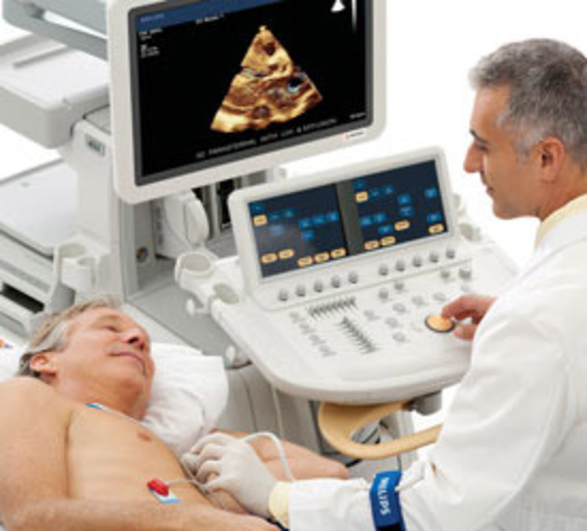
Transthoracic Echocardiogram Heart and Vascular
Echocardiography Imaging Techniques - NCBI BookshelfThis book chapter provides an overview of the basic principles and applications of echocardiography imaging techniques, including two-dimensional, three-dimensional, Doppler, and strain imaging. It also discusses the advantages and limitations of each technique, as well as the common artifacts and pitfalls that may occur. This book chapter is.
Echocardiography (ultrasound). Parker University
Biaya echo jantung untuk bayi dan anak-anak berkisar Rp500 ribuan sekali tes. Sedangkan echo jantung untuk orang dewasa berkisar di angka ratusan ribu hingga jutaan rupiah. Berikut daftar biaya echo jantung di sejumlah rumah sakit pada 2022. Biaya Pemeriksaan Echo (Ekokardiografi) Jantung - (www.heart.org)
Cardiac Radiology 2020 Lab
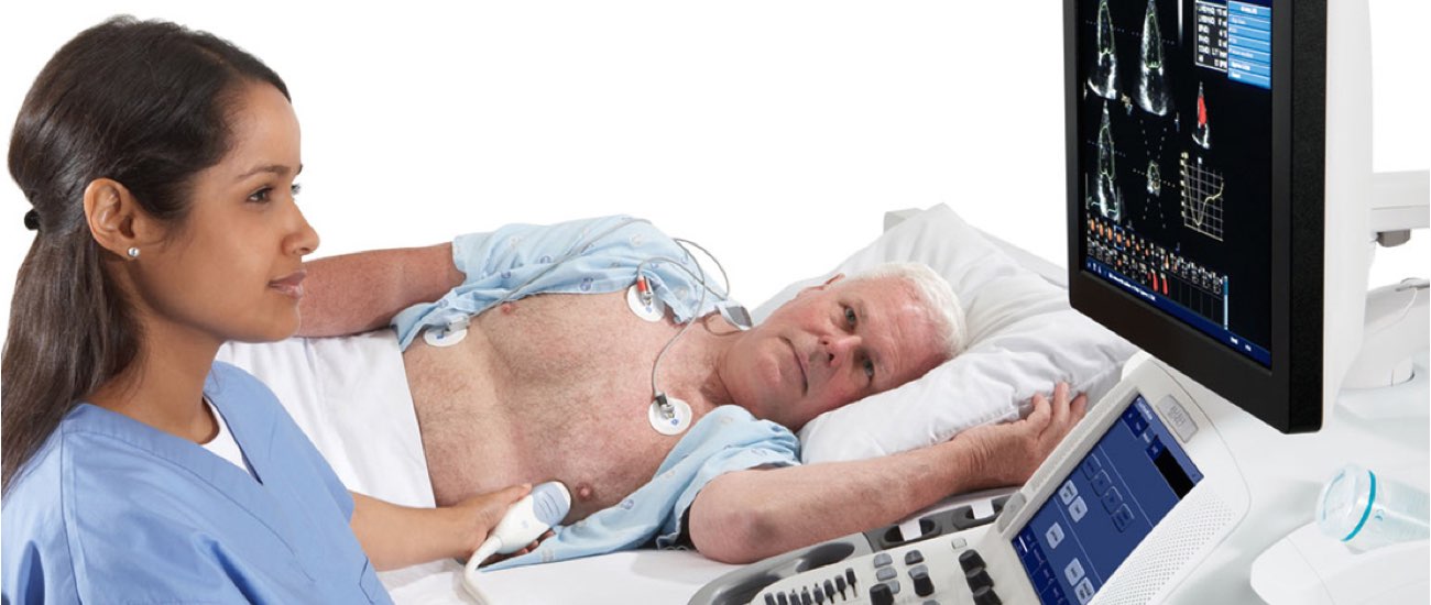
An echocardiogram is an ultrasound test that checks the structure and function of your heart. An echo can diagnose a range of conditions including cardiomyopathy and valve disease. There are several types of echo tests, including transthoracic and transesophageal. Talk with your provider about the type that's best for you.

An Atlas to Explore the Current Use of Echocardiography in Clinical Medicine Thoracic Key
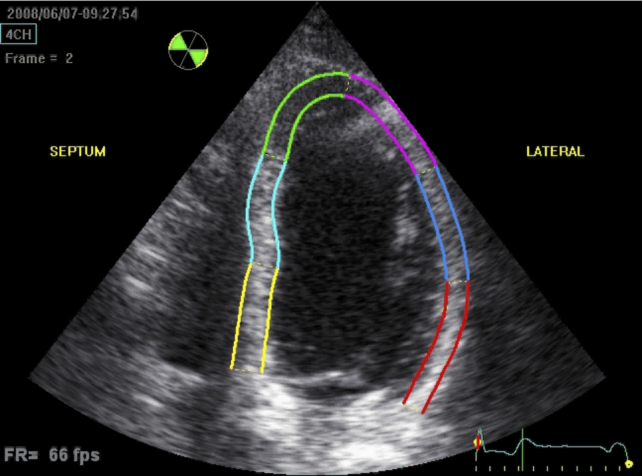
Echocardiography is the use of ultrasound to evaluate the structural components of the heart in a minimally invasive strategy. Although, prior to the invention of today's routinely used 2-dimensional echocardiography, there was motion-based (M-mode) echocardiography. In 1953, Inge Edler, regarded as the father of echocardiography, first described M-mode technology, which began the era of.
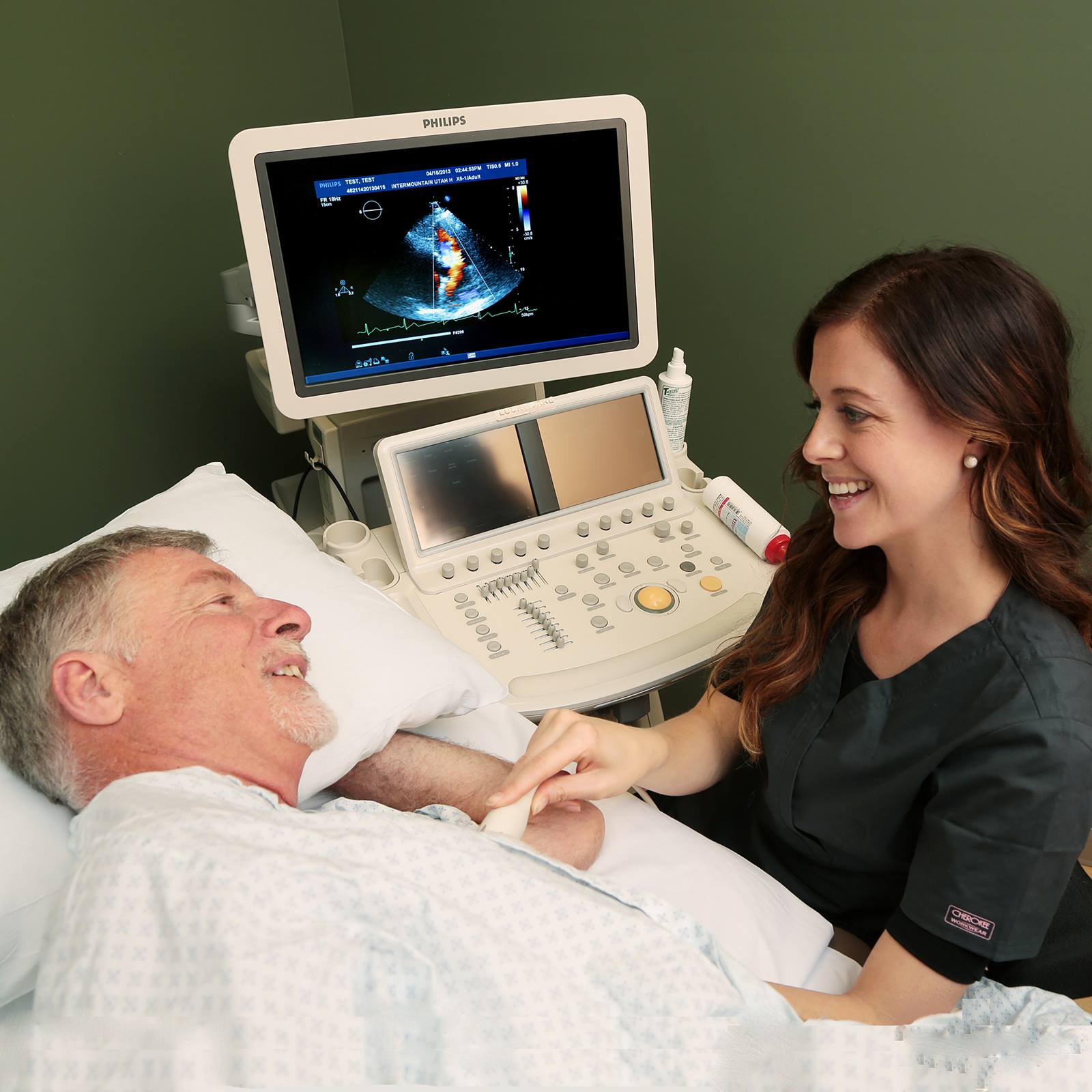
Echocardiogram Heart Care Intermountain Healthcare
Biaya untuk melakukan ekokardiografi bervariasi, tergantung dari jenis ekokardiografi dan rumah sakit mana yang menyelenggarakannya. Di rumah sakit swasta di Indonesia, biaya pemeriksaan ini bisa dimulai dari Rp. 400.000 hingga lebih dari Rp. 1.400.000. Dianjurkan untuk mempersiapkan dana lebih guna kebutuhan tambahan yang tidak terduga, yaitu.

How to use intracardiac echocardiography for atrial fibrillation ablation procedures Heart Rhythm
Echocardiography. Echocardiography uses ultrasound waves to produce an image of the heart, the heart valves, and the great vessels. It helps assess heart wall thickness (eg, in hypertrophy or atrophy) and motion and provides information about ischemia and infarction. It can be used to assess systolic function as well as diastolic filling.
Echocardiogram Definition, Uses, procedure & Side effects Irving Radiology
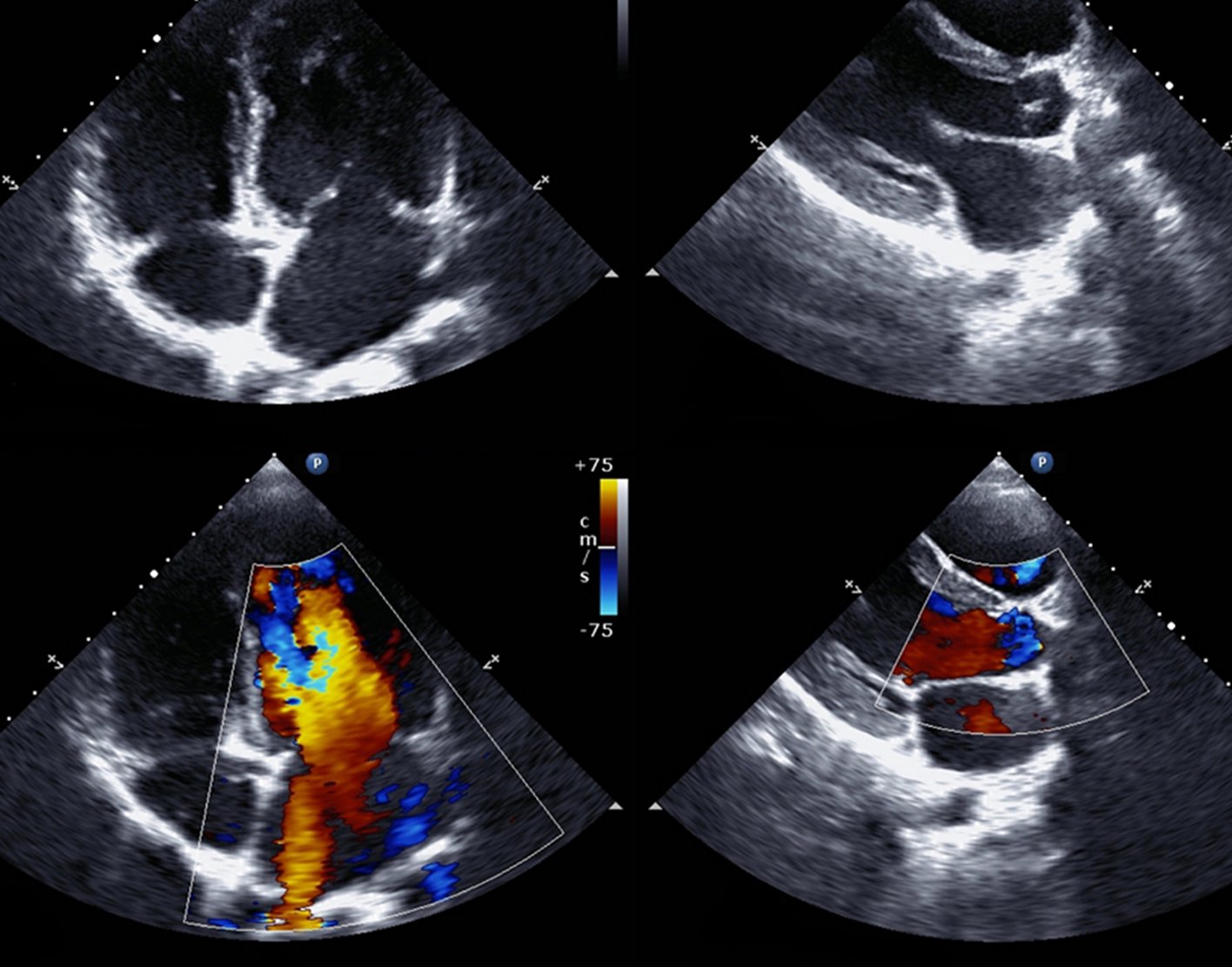
Echo is the cheapest and least invasive method available for screening cardiac anatomy. Generalists most commonly request an echo to assess left ventricular (LV) dysfunction, to rule out the heart as a thromboembolic source, and to characterize murmurs. The approximate normal values for various cardiac structures are described in Table 1.
Echocardiography — Cardiology Institute
Oversight, Review, and Final Edits by Vi Dinh (POCUS 101 Editor). A bedside Cardiac Ultrasound or Echocardiogram is a quick Point of Care Ultrasound (POCUS) that allows you to visualize and evaluate how the heart is functioning. In addition, bedside echocardiography also allows you to evaluate hemodynamic changes and pathological heart diseases.
What is an echocardiogram? Uses, procedure, and results
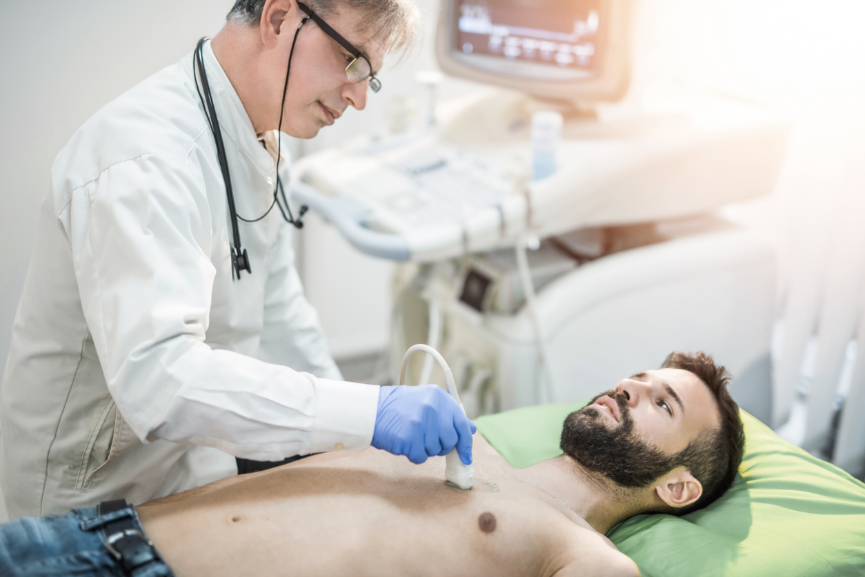
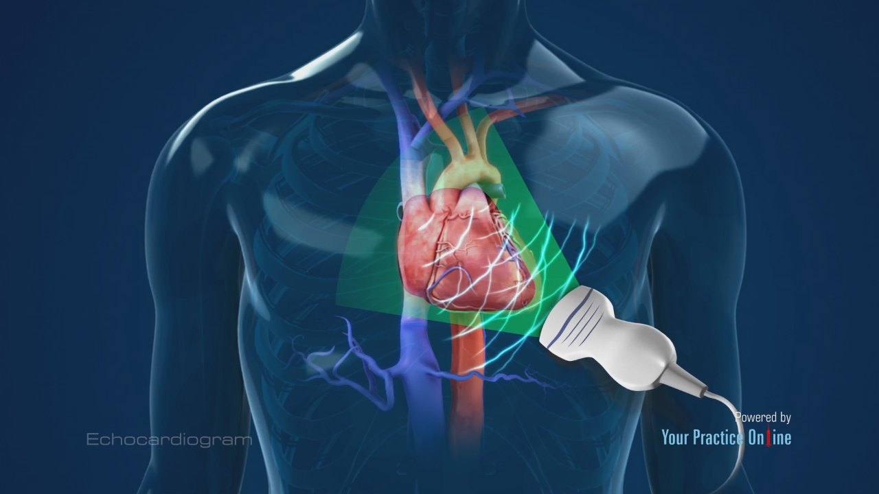
Echocardiogram. An echocardiogram is a noninvasive (the skin is not pierced) procedure used to assess the heart's function and structures. During the procedure, a transducer (like a microphone) sends out sound waves at a frequency too high to be heard. When the transducer is placed on the chest at certain locations and angles, the sound waves.
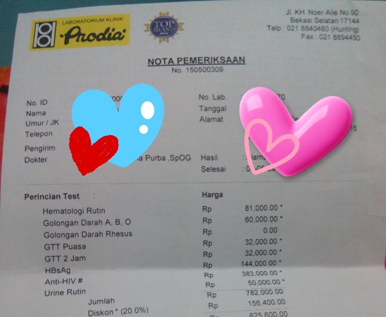
Biaya Echo Jantung Di Prodia Terbaru
Echocardiography uses ultrasound waves to produce an image of the heart, the heart valves, and the great vessels. It helps assess heart wall thickness (eg, in hypertrophy or atrophy) and motion and provides information about ischemia and infarction. It can be used to assess systolic function as well as diastolic filling patterns of the left.